Understanding Airborne Fibre Analysis: Phase Contrast Microscopy vs. Transmission Electron Microscopy
When it comes to analyzing airborne fibre concentrations—particularly in the context of asbestos monitoring—two prominent techniques are often employed: Phase Contrast Microscopy (PCM) and Transmission Electron Microscopy (TEM). While both methods strive to quantify airborne fibres, they each offer unique advantages and are suited for different scenarios.
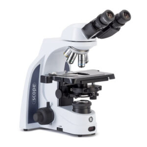
Phase Contrast Microscopy (PCM)
PCM is the primary method used for assessing “respirable” airborne fibres during standard asbestos monitoring. It employs the Membrane Filter Method (MFM), which estimates the concentration of airborne fibres over a specified timeframe. However, this approach is not without its challenges.
One significant drawback of PCM is the uncertainty that can arise from various factors. Field sampling errors, such as variations in pump flow rates and sampling locations, can greatly impact results. Additionally, high-density dust clouds can skew the data collected. In the laboratory, issues like microscope calibration and the experience level of the analyst further complicate accuracy. Notably, PCM cannot distinguish between asbestos and non-asbestos fibres when they meet the criteria for “respirable fibres.”
This limitation poses challenges in environments rich in synthetic mineral fibres or organic materials, such as commercial laundries or construction sites. In these settings, non-asbestos fibres may inflate fibre counts, potentially leading to regulatory interventions and operational delays. Therefore, ensuring that both field personnel and laboratory analysts are highly trained is crucial for obtaining reliable results.
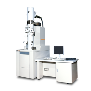
Transmission Electron Microscopy (TEM)
In situations where greater precision is required, especially for identifying specific types of airborne fibres, TEM emerges as the preferred method. This advanced microscopy technique offers extremely high magnification, enabling detailed observation of fibre morphology and mineral composition. Unlike PCM, which utilizes optical methods, TEM relies on electron microscopy, providing a more in-depth analysis.
Using the same filters from MFM, the TEM process involves several steps. The filters are processed, and the contents are placed onto a microscopy grid, often coated with carbon or gold to enhance visualization. This method allows for the characterization of fibres, offering insights into their specific types rather than simply providing a blanket count of all fibres present.
Conclusion
Alpha Analytics is actively collaborating with international companies to introduce Transmission Electron Microscopy (TEM) analysis to New Zealand. This partnership aims to enhance the capabilities of local air quality monitoring by providing access to this advanced technology. By bringing TEM to New Zealand, we seek to improve the accuracy and specificity of airborne fibre analysis, ensuring safer environments for all. Our commitment to innovation and collaboration positions us to lead the way in asbestos monitoring and contribute to more effective regulatory compliance.
For more information about this initiative, please contact Alpha Analytics Limited.